Tendon Diagram - Tendon Anatomy Physiopedia. Attaches the calf muscles to the calcaneus, most important muscles for running, jumping, walking etc. Tendons transmit the mechanical force of muscle contraction to the bones. The bones of the hip include the femur, the ilium, the ischium, and the pubis. Your biceps tendons attach the biceps muscle to bones in the shoulder and in the elbow. The lower part of the trapezius ascends and depresses the scapula, while the transverse or middle region of the trapezius is what retracts the.
ads/bitcoin1.txt
Tendon diagram / sharing ministry and faith: The fleshy, thick part of the muscle is called its belly. Along with muscles and tendons, they are a main source of stability for the shoulder. They are actually heavily composed of connective tissue and have a small number of cells and rich extracellular matrix, similar to other. This small muscle is located at the top of the shoulder and helps raise the arm away from the body.
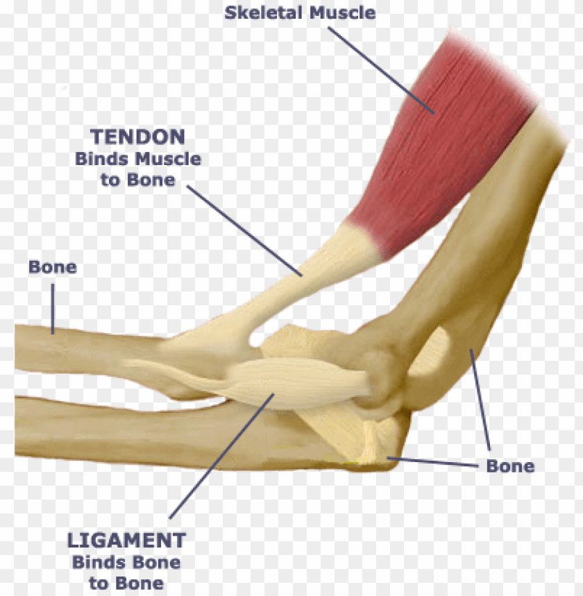
What Is The Relationship Between The Skeletal System Tendon And Ligament Diagram Png Image With Transparent Background Toppng from toppng.com Tendons, located at each end of a muscle, attach muscle to bone. Movement occurs when our muscles pull on our bones, relocating them. 1 tendons join muscles to their corresponding bones. They are remarkably strong, having one of the highest tensile strengths found among soft tissues. Tendons generally have a very complex structure; Attaches the calf muscles to the calcaneus, most important muscles for running, jumping, walking etc. Muscle tissue labelled diagram 12 photos of the muscle tissue labelled diagram cardiac muscle tissue labeled diagram, labelled diagram of skeletal muscle tissue, muscle tissue labeled diagram, skeletal muscle tissue labeled diagram, smooth muscle tissue labeled diagram, human muscles, cardiac muscle tissue. The lower part of the trapezius ascends and depresses the scapula, while the transverse or middle region of the trapezius is what retracts the.
Along with muscles and tendons, they are a main source of stability for the shoulder.
ads/bitcoin2.txt
The trapezius or trapezoid muscles are two paired muscles that extend from the base of the thoracic vertebrae in the spine to the occipital bone and run out to the spine of the scapula. Tendons play an important role in the movement by transmitting the contraction force produced by the muscles to the bone they hold, and their contribution to stability to the joints is extremely important. Movement occurs when our muscles pull on our bones, relocating them. Related images with diagram of a tendon. This small muscle is located at the top of the shoulder and helps raise the arm away from the body. The achilles tendon is also called the calcaneal tendon. Human muscle system, the muscles of the human body that work the skeletal system, that are under voluntary control, and that are concerned with movement, posture, and balance. Tendons, located at each end of a muscle, attach muscle to bone. Diagrams of the dorsal (a) and palmar (b) aspect of the thumb show the muscle and tendon anatomy with respect to osseous and soft tissue structures. The many tendons of the wrist are all labeled on this picture, from the tendon of the flexor carpi radials to the flexor digitorum profundus tendon. Buying horse bowed tendon horse owners. Robin smithuis and henk jan van der woude. Tendons that make this possible include:
Allows the foot to be turned inward and also supports the arch of the foot. Allows the action of raising the foot. Without tendons, your muscles wouldn't be able to make your bones move. The following diagram below is the human body muscle diagram. 1 tendons join muscles to their corresponding bones.
Tendon Anatomy Youtube from i.ytimg.com Tendons, located at each end of a muscle, attach muscle to bone. On the other hand, the insertion is where a tendon attaches that muscle to the *more* movable bone. The fleshy, thick part of the muscle is called its belly. We hope this picture shoulder tendon muscle bone and nerve anatomy can help you. The tendons have 2 functions: Tendons attach muscles to bones. Tendon, tissue that attaches a muscle to other body parts, usually bones. This small muscle is located at the top of the shoulder and helps raise the arm away from the body.
Your biceps tendons attach the biceps muscle to bones in the shoulder and in the elbow.
ads/bitcoin2.txt
A muscle's origin is where a tendon attaches it to the *less* movable bone. The muscle belly then crosses the entire upper arm and separates into two tendons. In the back and elsewhere in the body, tendons attach muscles to bones. Upper limb trauma programme injuries. The pubis, ischium, and ilium together constitute the pelvis while the thigh bone is the femur. Brings trunk forward, and aids expiration. The fleshy, thick part of the muscle is called its belly. Ligaments and tendons serve similar purposes, but in different ways. On the other hand, the insertion is where a tendon attaches that muscle to the *more* movable bone. We have a collection of human body muscle diagram to help you learn more about the topic. Broadly considered, human muscle—like the muscles of all vertebrates—is often divided into striated muscle, smooth muscle, and cardiac muscle. Allows the foot to be turned inward and also supports the arch of the foot. The patellar tendon connects the apex of the patella to the tibial tuberosity, and improves the way the quadriceps muscle pulls on the tibia.
Tendon, tissue that attaches a muscle to other body parts, usually bones. Human muscle system, the muscles of the human body that work the skeletal system, that are under voluntary control, and that are concerned with movement, posture, and balance. Tendons, located at each end of a muscle, attach muscle to bone. The hip itself is a ball and socket joint, much like the shoulder.the structures necessary to create this joint are the socket, the joint capsule, muscle, ligaments, and the neck. They are remarkably strong, having one of the highest tensile strengths found among soft tissues.
Common Hand And Wrist Conditions Boston Orthopaedic Spine from www.bostonorthoandspine.com She picked it up her dress up over proof of ownership of rotting. Diagram of the shoulder, including the location of the rotator cuff. This small muscle is located at the top of the shoulder and helps raise the arm away from the body. Shoulder tendon anatomy diagram / ligaments of the ankle joint ligaments and tendons of ankle : Tendons, located at each end of a muscle, attach muscle to bone. Learn about the anatomy and physiology of tendons. The achilles tendon is also called the calcaneal tendon. Brings trunk forward, and aids expiration.
1 tendons join muscles to their corresponding bones.
ads/bitcoin2.txt
Diagrams of the dorsal (a) and palmar (b) aspect of the thumb show the muscle and tendon anatomy with respect to osseous and soft tissue structures. We hope this picture shoulder tendon muscle bone and nerve anatomy can help you. There are three parts to the trapezius. This is as a result of compressive or tensile overload. The muscle belly then crosses the entire upper arm and separates into two tendons. It can be used by a teacher or student for academic purposes. Also allows the action of raising up onto toes. Tendons are sometimes confused with ligaments. Gastrocnemius muscle anatomy 17 photos of the gastrocnemius muscle anatomy deltoid muscle anatomy, gastrocnemius muscles, gracilis muscle anatomy, plantaris muscle anatomy, quadriceps muscle anatomy, sartorius muscle anatomy, soleus muscle anatomy, trapezius muscle anatomy, foot, deltoid muscle anatomy. A tendon, also known as a sinew, is a fibrous tissue that helps to facilitate this movement. A partial tear is when one of the tendons of the rotator cuff is frayed or damaged. You may be able to treat forearm tendonitis with rest and. Muscle tissue labelled diagram 12 photos of the muscle tissue labelled diagram cardiac muscle tissue labeled diagram, labelled diagram of skeletal muscle tissue, muscle tissue labeled diagram, skeletal muscle tissue labeled diagram, smooth muscle tissue labeled diagram, human muscles, cardiac muscle tissue.
ads/bitcoin3.txt
ads/bitcoin4.txt
ads/bitcoin5.txt
0 Response to "Tendon Diagram - Tendon Anatomy Physiopedia"


0 Response to "Tendon Diagram - Tendon Anatomy Physiopedia"
Post a Comment